Advanced Microelectronics System for Neural Prosthesis and Organ Centric Disease Treatment
Research Motivations
- Electroceuticals
- When drugs can't coax cells in the pancreas to produce insulin, or loosen arteries to reduce blood pressure, a well-placed jolt of electricity might do the trick. Spurred by decades of success with pacemakers and cochlear implants, and by advances in miniaturized technology, interest is surging in 'electroceuticals' - bioelectronic implants that stimulate nerves to treat disease. More about electroceuticals: news
- BRAIN Initiative
- The BRAIN Initiative is a proposed collaborative research initiative announced by the Obama administration on April 2, 2013, with the goal of mapping the activity of every neuron in the human brain. Current technology allows researchers to observe about 100 neurons in real time. Researchers argue funding for the BRAIN Initiative would help scale up new technology towards real-time recordings of approximately one million neurons in a human brain in about 15 years. More about BRAIN Initiative: wiki, discussion
Neural Technology Chip Gallery by TSS Group
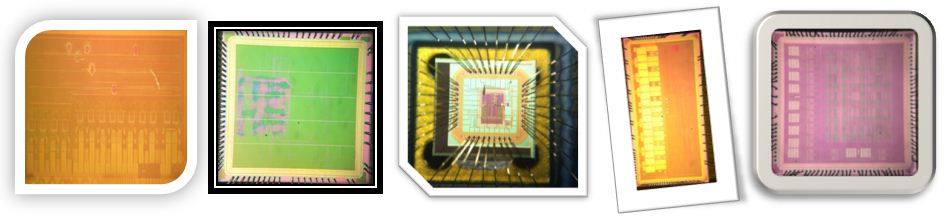
We have been working on microelectronics for years and developed critical IPs to build up future platform technology, including
- an innovative integrated, low power peripheral neural recorder that can possibly survive the large artifacts (up to 1V@4Hz) from the peripheral nerve recording environment and maintains extremely low noise (1µV) to resolve axonal signals.
- a set of neural signal processing algorithms implemented in integrated circuits, enabling a new term called " in-implant computing". Compared with other approaches, our signal processing features a reliable and automatic operation supplement by cutting edge VLSI circuits implementation. Our IPs have been verified in behavior monkey experiments and achieved impressive accuracy.
- an innovative design to achieve simultaneous neural stimulation (current stimuli up to Vdd) and spike recording. We are porting the design into a high voltage (60V/5V/1.8V) Bipolar-CMOS-DMOS process. This is a technology for closed-loop stimulation in Electroceuticals.
- an innovative system platform named Neuronix, where each recorder (a few hundred µm size) can simultaneously acquire data from multiple electrodes. So far we have demonstrated one Neuronix channel to simultaneously record 2 electrodes, while our ultimate goal is to have a sub-mm sized device to record 1,000 channels. Once succeed, it is a powerful technology that is in line with the BRAIN Initiative.
- a high density, high intensity, high frame rate, flexible patterned optical stimulator for in-vitro neuroscience experiments. We are conducting in-vitro stimulation experiments with hippocampus tissues and electrically recording through MEA.
An Integrated, Low Power, Low Noise Peripheral Nerve Recorder
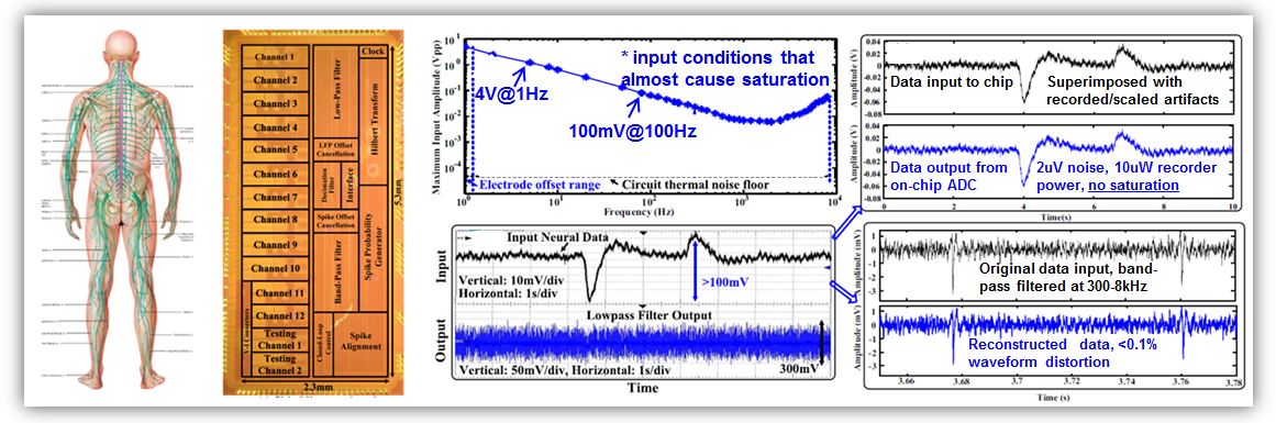
We have designed, fabricated, and tested a recorder for peripheral nerve recording. The circuits are based on an innovative architecture, achieving high amplifier input impedance (3pF), low noise operation (2µV@300-8kHz or 1µV@700-4kHz), and an ultra wide recording dynamic range, i.e., we have tested 4V sine input at 1Hz (motion artifacts), 100mV sine input at 100Hz (EMG artifacts), both not causing saturation. Such wide dynamic range is to accommodate artifacts that frequently appear in peripheral nerve recordings and prevent signals being masked.
Unlike commonly used peripheral nerve amplifiers that are bulky (discrete circuits board) and power hungry (100mW per amplifier), our recorder is fully integrated and consumes less than 10µW per channel (including ADC and digital blocks). In addition, our recorder does not suffer from high pass filter noise (a dominant noise source in peripheral nerve recording) and ensures high quality recording regardless of small/large electrodes, with/without artifacts. We have finished system development and device characterization. In-vivo peripheral nerve recording experiments are ongoing.
In-Implant Computing - Information Decoding
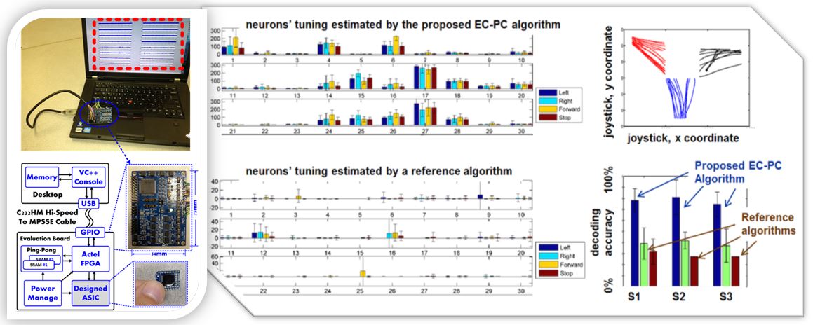
We have invented and applied the EC-PC detection algorithm to monkey experiments and discovered that it can lead to large improvement in decoding accuracy. In the experiment, a monkey was asked to use a joystick to control the mobile platform it was sitting on and direct the platform towards a caretaker, which holds a juice award. A 100-channel microelectrodes array was implemented on the motor area of the monkey's brain. The upper right figure shows the trajectory of the joystick movement. The bottom right figure shows that the EC-PC algorithm almost doubled the decoding accuracy compared with Non-linear Energy Operator (NEO) and simple threshold spike detection, across 3 sessions of experiments. In addition, our EC-PC algorithm does not require any parameter tuning (fully automatic and parameter learnt on-the-fly). From neurons' tuning, we can see that firing rate estimated based on EC-PC spike detection is more consistent (with lower intra-class variance), and easier to classify (with larger inter-class mean difference). We have implemented our algorithm into a digital ASIC chip integrated with analog frontend. The left figure shows an earlier version of the EC-PC ASIC chip, which consumes 80-100µW for processing on-channel neural data continuously. We have spent major efforts to optimize our algorithm, increase circuit efficiency, and integrate advanced circuits IPs. A future version under development only consumes 3µW per channel power. Once done, we will have extremely low power, yet reliable neural signal processing ASIC for information decoding.
In-Implant Computing - Data Compression and Chip Optimization
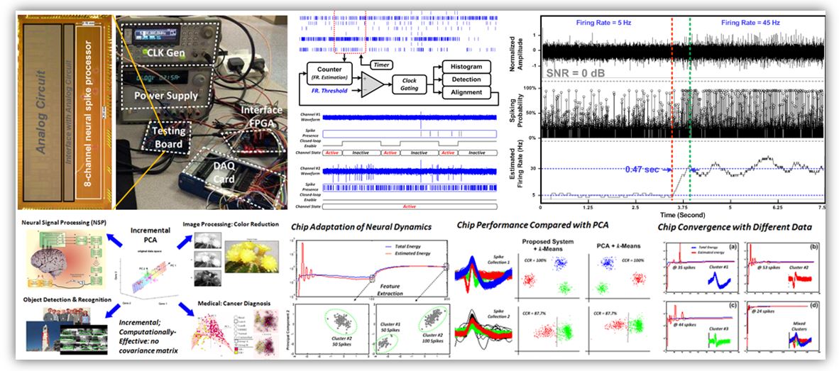
We have accumulated a set of algorithm ASICs for processing neural data, including noise reduction, interference removal, artifact removal, spike detection, feature extraction, classification, and firing rate estimation. These algorithms are to be implemented inside the implant for real-time, continuous operation. Our motivation is two-fold, one is to achieve a substantial amount of data rate reduction thus allowing wireless data communication between the implant and an external piece, i.e., a wearable. For example, our algorithm can compress the data by 100-10,000 times (an exact compression ratio is data dependent) in measurement without loss of information. The second motivation is to adaptively configurable circuit and achieve lower power operation. For example, in a recent recorder chip, a closed-loop control mechanism has been made possible by estimating firing rates based on alignment results and turning on/off channels individually and automatically: if there are no/little spikes, the channel is configured into a low frequency mode for recording field potentials only - this gives 100 times power reduction. When spikes are detected, the channel is configured back to full bandwidth. Our recorder then only consumes the just needed power according to signal characteristics.
Nonstopped Neural Recording during Stimulation
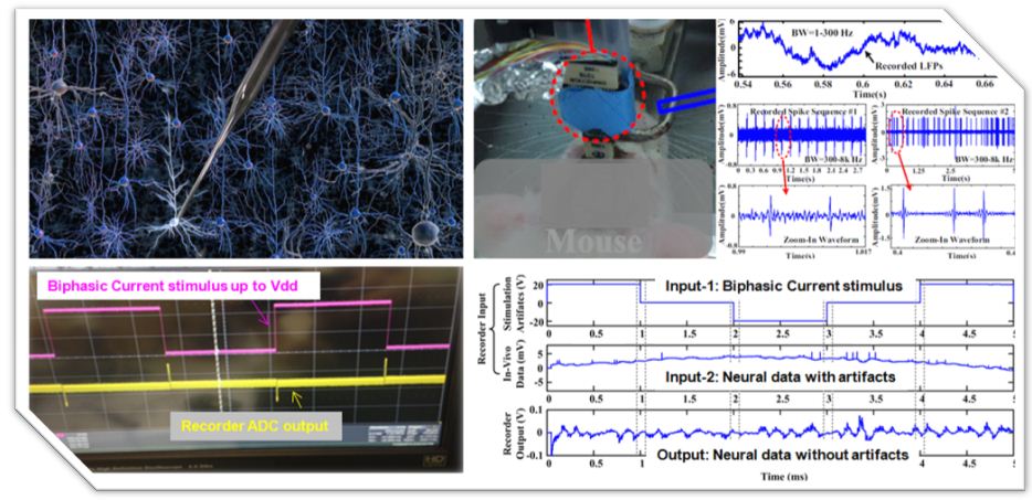
Functional electrical stimulation has been used as a treatment option for many diseases. Despite major success in improving patient life quality and treatment of intractable diseases, there could be substantial side effects. To benefit patients and increase the market size, it is important to target unmet clinical needs and make improvements on stimulation selectivity, safety, reliability, size, power consumption, and endurance. In this work, we propose a closed-loop neural stimulator, which allows recording direct neuronal responses during or after stimulation. With this information, it is possible to adjust stimulation pulse width, amplitude, and duty cycle for optimal stimulation outcome, i.e., less tissue damage, improved spatial selectivity, less power consumption, and individualized stimulation protocols. As shown in the bottom left figure, the purple trace is a measured biphasic current stimulus up to Vdd. With a conventional amplifier, the biphasic current pulse saturates the recorder, where it takes about tens of ms to a few hundred ms to recover. The yellow trace is the measured waveform from the recorder output (ADC output), where saturation only appears at the stimulus transition edges. The duration of the un-recordable period can be controlled by adjusting power: with 200µA power budget allocated to the amplifier, the un-recordable period is about 25µs. The missing sample can be interpolated thus it allows nonstopped recording during stimulation.
Neuronix - A Future Technology Towards Massive Neural Recording
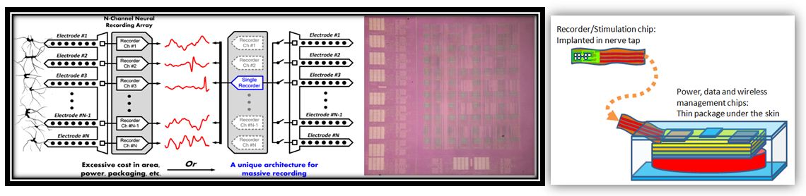
Neuronix is an innovative system platform, where each recorder (a few hundred µm size) can simultaneously acquire data from multiple electrodes. So far we have demonstrated one Neuronix channel to simultaneously record 2 electrodes, while our ultimate goal is to have a sub-mm sized device to record 1,000 channels. In collaboration with material scientists with fabrication facilities, we are building up an implant technology of unprecedented recording density that is in line with the BRAIN Initiative. In collaboration with neuroscientists, neural engineers, and physicists, we are developing a new neural interface for peripheral nerve recording and stimulation of much improved recording/stimulation selectivity. This is then a powerful technology for Electroceuticals. More information is available upon request.
An Optical Stimulator for In-Vitro Neuroscience Experiments
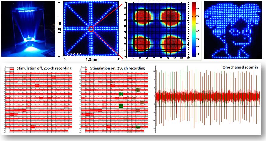
Recent optical stimulation technologies allow improved selectivity and have been widely used in neuroscience research. This work presents an optical stimulator based on mm sized, bright LEDs. It has 1024 channels and can produce flexible stimulation patterns in each frame, refreshed at above 50 Hz. To increase the light intensity, each LED has an optical package that directs the light into a small angle. To ensure that the light of each LED can reach the lens, the LEDs have been specially placed and oriented towards the lens. With these efforts, the achieved power efficiency (defined as the amount of LED light power passing through the lens divided by the LED total power consumption) is 0.00005. In our current prototype, an individual LED unit can source 60mW electrical power, where the induced irradiance on neural tissues is 6 mW/mm2 integrating from 460nm to 480nm. The light spot is tunable in size from 18 µm to 40 µm with an extra 5-10 µm separation for isolating two adjacent spots. Through both bench-top measurement and finite element simulation, we found that the cross channel interference is below 10%. The developed optical stimulator is used as a tool in neuroscience experiments for studying biological memory.